
FROM MY PT SCHOOL NOTES · NEUROLOGY · UNIT 7 · CHAPTER 25
How does vestibular examination inform rehabilitation?
Vestibular rehabilitation is not one generic set of head or eye exercises. Students need to link the anatomy, clinical syndrome and examination to the specific problem.
Examination and clinical reasoning
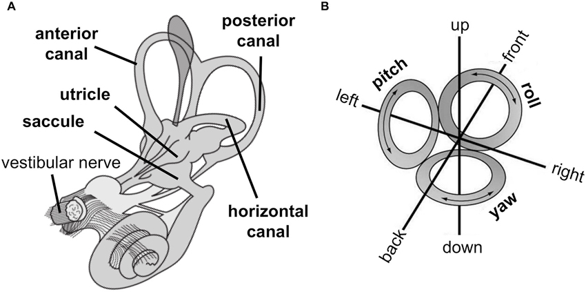
Understand the vestibular contribution
Semicircular canals detect angular head motion; the utricle and saccule contribute to linear acceleration and head orientation. Vestibulo-ocular reflex pathways help stabilize vision while the head moves. Balance depends on interaction among vestibular, visual, somatosensory, cerebellar and musculoskeletal systems.
Symptoms may involve oscillopsia, dizziness, imbalance or motion sensitivity. Similar complaints can arise from cervical, migraine, medication, visual or neurological causes; the diagnosis is not made by symptom intensity alone.
Separate positional disorders from hypofunction
For posterior canal BPPV, the AAO-HNS guideline supports skilled positional testing and canalith repositioning in the appropriate presentation. Avoid routinely prescribing Epley for all dizziness or outdated post-maneuver restrictions.
For confirmed vestibular hypofunction, tests of gaze stability, balance and walking can identify functional limitations. Patient-reported symptom measures are valuable but are not standalone diagnostic tests. Use official scoring instructions and evaluate safety before head/neck maneuvers.
Match intervention to target
The 2022 ANPT vestibular hypofunction guideline supports appropriately selected gaze-stabilization exercises with head movement and balance/gait rehabilitation. Smooth pursuits or saccades in isolation should not replace gaze-stabilization work for hypofunction.
Set dose and progression according to condition, symptoms, comorbidity and functional demands. BPPV repositioning, VOR adaptation, habituation and functional balance training have different purposes; avoid trying to provoke maximum symptoms as an end in itself.
Quick distinctions
| BPPV | Canal-specific positional assessment and repositioning |
| Vestibular hypofunction | Gaze stability with head movement, balance and walking |
| Atypical findings | Refer or seek specialist assessment before selecting maneuvers |
Clinical reasoning case
A patient with chronic oscillopsia while walking is given smooth-pursuit eye tracking with a still head as the only treatment.
Reasoning: That alone does not directly train the impaired head-motion gaze-stability response; after appropriate assessment, targeted VOR and functional balance/gait work is more relevant.
Check your understanding
How is BPPV repositioning different from VOR adaptation training?
Original uploaded sources: 13 Vestibular System.pdf; 6 Vestibular Disorders.pdf; Cawthorne Cooksey Exercises.pdf; Dizziness Handicap Inventory.pdf (not reproduced).
Historical note: Compare historic exercise handouts with the 2017 BPPV and 2022 vestibular hypofunction guidelines.
Educational material, not a patient-specific diagnostic or emergency-care protocol.


