
← Diagnostic Imaging class notes
FROM MY PT SCHOOL NOTES · DIAGNOSTIC IMAGING · UNIT 2 · CHAPTER 08
Elbow
Review the relevant anatomy and imaging question before deciding what the finding may mean for rehabilitation.
Key imaging concepts
- Examine AP and lateral views for bony continuity, radial head/capitellar alignment, joint congruence and effusion signs.
- The fat-pad signs on lateral imaging may signal an intra-articular injury after trauma even if a fracture line is subtle.
- Postoperative implants and prior surgery alter landmarks; understand the procedure before comparing with typical anatomy.
PT clinical connection
After a fall, radial head tenderness, restricted rotation or unexpected swelling requires appropriate medical assessment.
High-yield distinction
Post-traumatic fat pad sign: The visible posterior fat pad can indicate effusion and possible occult intra-articular injury; correlate with examination and referral pathway. VERIFIED REAL-IMAGE REFERENCES (SOURCE LINKS ONLY) • Normal elbow AP · CC0 1.0 · normal view; cannot illustrate effusion • Fat-pad sign · CC BY-SA 3.0 · same sign; share-alike obligations must be handled
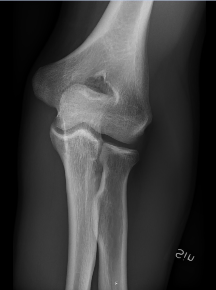
Course source: 2 Elbow.pdf; 2 Elbow Updated.pptx. These are educational notes, not image reading certification or a substitute for an official report. Imaging orders and interpretation rights vary with professional scope and state law; image examples must be clinically verified and properly licensed.
← Unit 2 · ← Previous · Next →


