
← Diagnostic Imaging class notes
FROM MY PT SCHOOL NOTES · DIAGNOSTIC IMAGING · UNIT 3 · CHAPTER 11
Knee
Review the relevant anatomy and imaging question before deciding what the finding may mean for rehabilitation.
Key imaging concepts
- AP, lateral and patellofemoral views may answer different questions. Assess joint space, tibial plateau, patellar position, cortical edges and effusion.
- A lateral plateau fracture can be subtle; high-energy trauma and inability to bear weight require appropriate assessment.
- MRI may be considered for meniscus, ligament or marrow questions when results will affect care, not merely because pain persists.
PT clinical connection
Apply validated acute trauma criteria appropriately; avoid using knee decision rules for chronic atraumatic symptoms.
High-yield distinction
View-dependent narrowing: Joint-space width depends on projection and loading. Do not compare non-weight-bearing views as if they were identical to weight-bearing films.
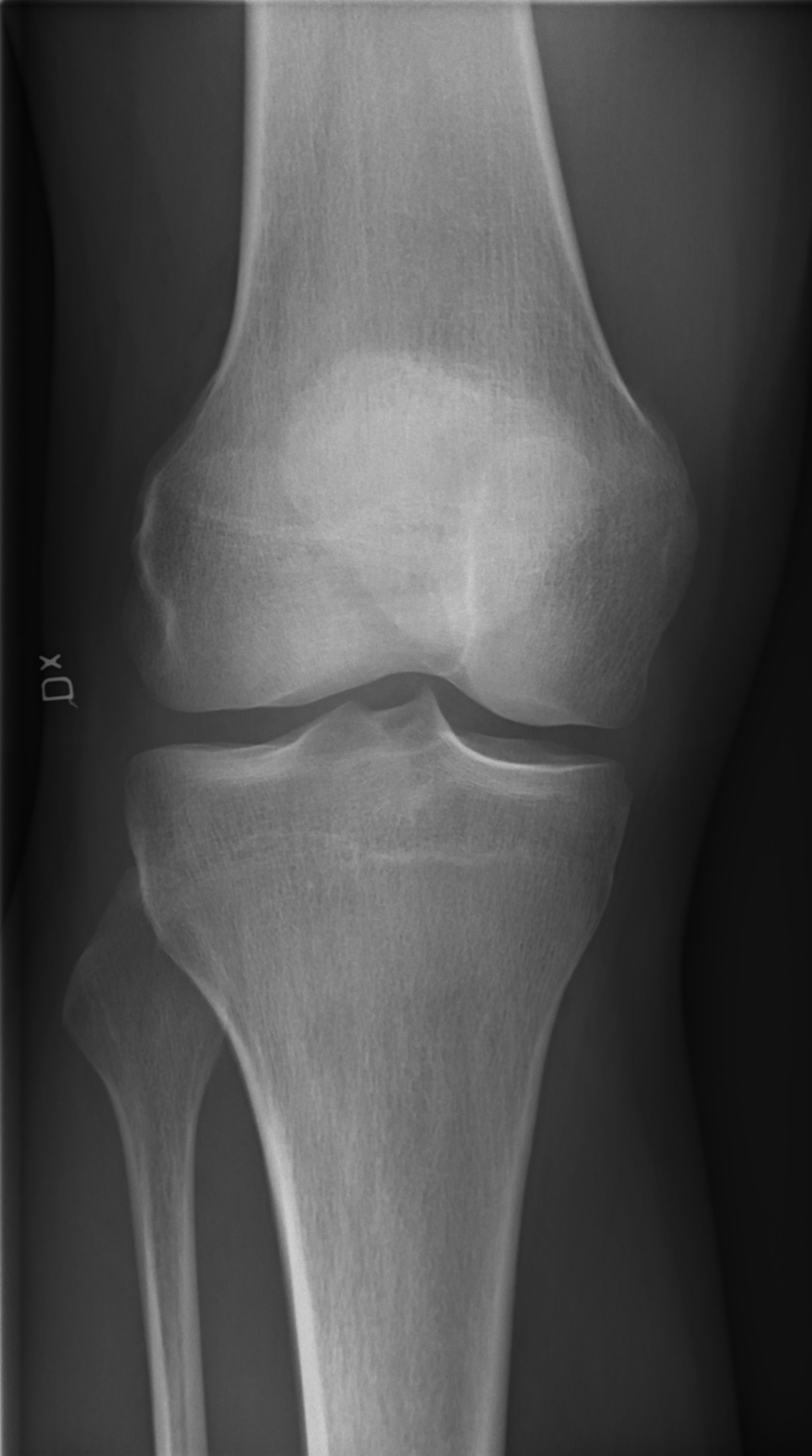
Course source: 5 Knee.pdf; 5 Cases for knee.pdf. These are educational notes, not image reading certification or a substitute for an official report. Imaging orders and interpretation rights vary with professional scope and state law; image examples must be clinically verified and properly licensed.
← Unit 3 · ← Previous · Next →


