
FROM MY PT SCHOOL NOTES · GENERAL MED / WOUND CARE · CHAPTER 13
What Has to Happen for a Wound to Heal?
Wound healing is a process involving barrier restoration, tissue formation and remodeling—not just the appearance of a scab.
Know the skin layers
Skin has three broad regions: the epidermis forms the outer barrier; the dermis contains vessels, connective tissue, nerves, hair follicles and glands; and the subcutaneous tissue provides padding and connects skin to deeper structures. The epidermis is avascular and depends on underlying tissues for nourishment. Damage depth affects the tissue’s capacity to regenerate and the potential for scarring.
Wound closure by intention: primary intention means the wound edges are approximated (such as a sutured incision); secondary intention involves filling an open defect through granulation, contraction and epithelialization; delayed primary or tertiary intention involves closure after a period of open management. These describe how a wound is closed, not a guarantee of normal healing.
Recognize phases of repair
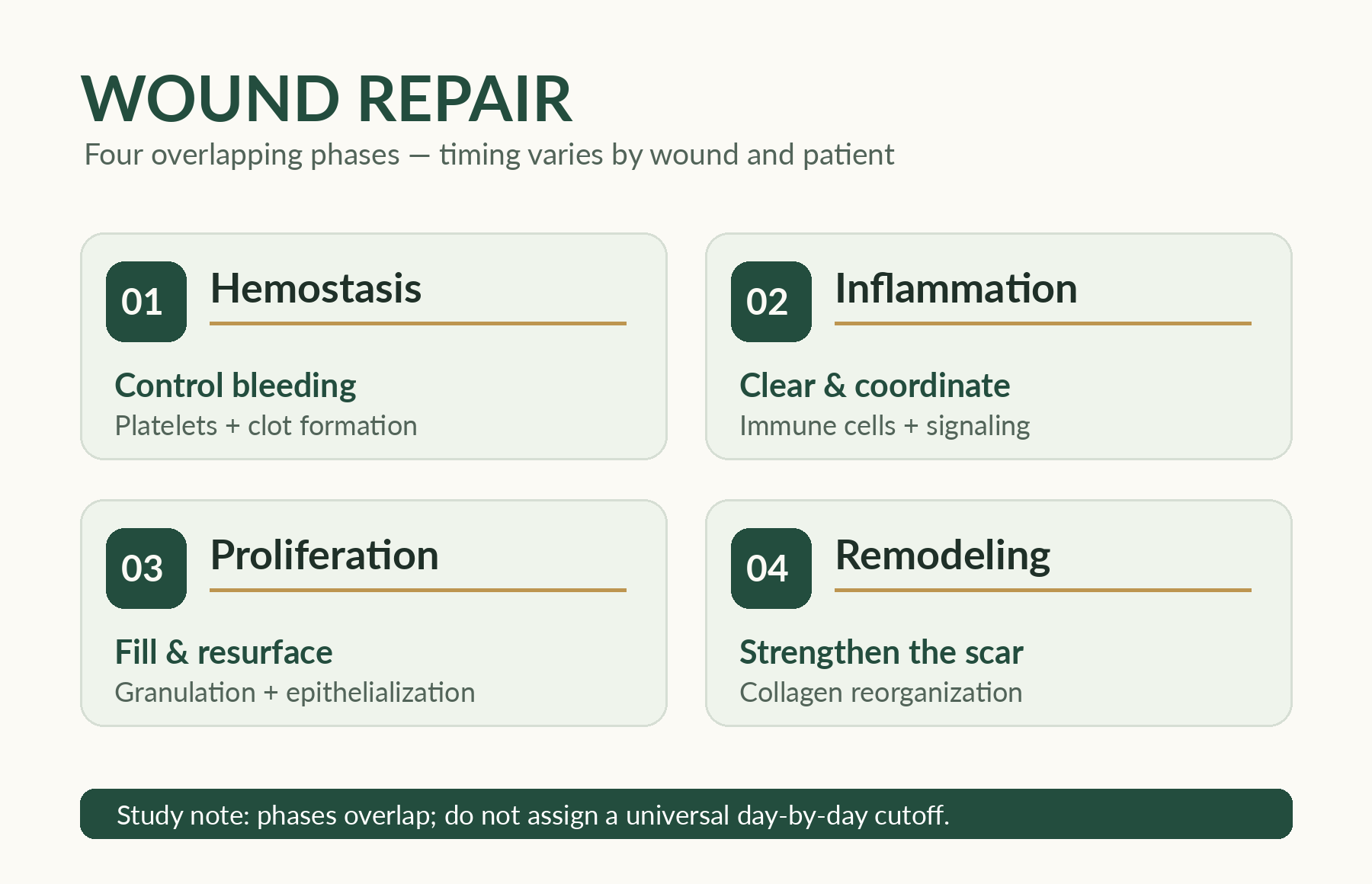
The extracellular matrix (ECM) helps explain why repair is more than simply forming new skin. Collagen contributes tensile strength; elastin contributes stretch and recoil; glycosaminoglycans help with tissue hydration and signaling; enzymes and growth factors help regulate matrix turnover. Too much breakdown relative to repair may contribute to a nonhealing wound.
Additional ECM notes: fibrin forms an early provisional scaffold. Growth factors (including platelet-derived growth factor, PDGF) and cytokines influence cell recruitment, proliferation and signaling. Fibroblasts lay down new matrix, while matrix metalloproteinases (MMPs) participate in breakdown and remodeling. A persistent imbalance between tissue breakdown and formation is one mechanism of impaired chronic-wound healing. Collagen becomes reorganized during scar maturation; a closed wound is not yet as strong as uninjured tissue.
Local factors: inadequate oxygen delivery or perfusion; excess pressure, friction or shear; foreign material; desiccation or maceration; excessive inflammatory burden; poor edge progression; repeated trauma; and unmanaged swelling. A chronic wound frequently has more than one active contributor.
Identify barriers to progression
The course organizes barriers to repair into local (perfusion, pressure and shear, moisture imbalance, inflammation and foreign material), systemic (chronic disease, impaired nutrition, smoking, immunosuppression and medications), and iatrogenic factors (overly aggressive cleansing, adhesive trauma, or unsuitable dressings). A granulating wound can be harmed by excessive intervention. Moist wound healing supports epithelial migration when excess drainage is controlled; too much moisture can macerate the surrounding skin.
Systemic factors: diabetes, peripheral arterial disease, venous disease, immune suppression, malnutrition risk, smoking, selected medications, major illness, reduced mobility and impaired self-care. Treatment burden also matters: dressing cost, availability, caregiver ability and adherence can affect outcomes.
Microbiology note: contamination means microorganisms are present; colonization means they persist or multiply without an established harmful host response; clinical infection means microorganisms are causing a damaging tissue/host response. Biofilms are organized microbial communities that can contribute to persistent wound problems. A positive culture, odor or color alone does not establish the diagnosis. Consider local signs, patient risk, changes in wound trajectory and systemic symptoms.
Check your reasoning: Can a wound appear less red while still healing poorly because of pressure or perfusion?
Yes. Evaluate the whole wound, tissue status, ongoing loading and systemic factors rather than one visible feature.
Make the clinical connection
Scenario: A sutured incision is closing edge-to-edge, while a deeper open wound is filling gradually with granulation tissue.
What is the next reasoning step?
Reasoning: The first generally illustrates healing by primary intention; the second illustrates secondary intention. Different tissue patterns and risk factors require different monitoring. Neither appearance alone proves successful healing.
Common mistake: Using one visible sign as a complete assessment of wound progress. Remember: Know the tissue and process you are observing.
Source: my General Medical / Wound Care class-note companion, wound section(s) 1 and 2. This is a conceptual educational lesson, not a wound assessment credential or a patient-specific treatment protocol. Dressing, staging and intervention decisions require appropriate training and current professional guidance.

