
FROM MY PT SCHOOL NOTES · WOUND CARE · CHAPTER 19
How Do You Distinguish Venous From Arterial Wound Patterns?
Two wounds on the lower leg can look similar but have different causes and treatment implications.
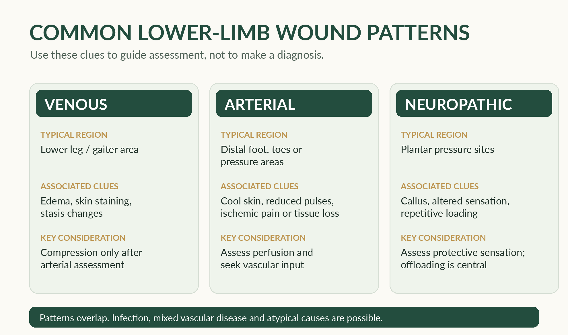
Look for perfusion and edema features
Venous disease: faulty valves, venous obstruction or impaired calf-muscle pumping can contribute to venous hypertension. Typical clinical clues may include lower-leg edema, skin darkening from hemosiderin, stasis dermatitis and shallow irregular ulcers often in the gaiter region. A location or color alone is not diagnostic.
Understand compression in context
Arterial disease: inadequate blood flow can present with exertional leg symptoms, rest pain, cool or poorly perfused tissues, diminished pulses and toe/foot ulcers with tissue loss. Some patients—especially those with diabetes—have mixed or atypical presentations. A small-looking ulcer can represent major ischemic risk.
Why compression matters: for appropriate venous leg ulcers, compression helps counter venous hypertension and edema and is a major treatment principle. Multi-layer wraps, short-stretch materials, stockings and an Unna boot differ in properties and practicality. Exercise can support calf-muscle pumping; elevation may help selected patients, but these do not replace vascular assessment.
Recognize mixed or concerning presentations
Unna boot class note: a zinc-impregnated, relatively inelastic wrap is one compression option for selected venous cases. The class lab emphasizes keeping it dry and checking dressing fit and skin tolerance. Assess perfusion, infection concerns, drainage, function and follow-up before use. Application is a trained procedure—not a general-purpose home-care recommendation.
Compression method comparison: the lecture distinguishes relatively inelastic short-stretch wraps, elastic long-stretch systems, multi-layer wraps, and stockings. They differ in resting and working pressure, mobility demands, tolerance and application requirements. Select a method only after adequate perfusion assessment and with a feasible follow-up plan.
Check your reasoning: Is compression automatically appropriate for every swollen leg wound?
No. Arterial perfusion, the patient’s medical status and the type and degree of compression must be assessed.
Make the clinical connection
Scenario: A swollen lower leg has an irregular superficial ulcer and skin discoloration, but the pedal pulses seem reduced and the person reports pain at rest.
What is the next reasoning step?
Reasoning: Venous features may be present, but arterial or mixed disease must be evaluated. Compression should not be started blindly from appearance or edema alone; seek the appropriate vascular assessment and clinician guidance.
Common mistake: Treating visible swelling as automatic proof compression is safe. Remember: Clarify blood supply and the overall wound cause before compression decisions.
Source: my General Medical / Wound Care school-notes companion, wound section(s) 8. This is for studying concepts and clinical reasoning. It does not authorize debridement, compression, dressing selection or burn care without qualified training, evaluation and current protocols.

