
FROM MY PT SCHOOL NOTES · NEUROLOGY · UNIT 2 · CHAPTER 06
How do motor, sensory, reflex and cranial nerve findings fit together?
A neurological examination is an organized search for patterns, not an attempt to check every nerve in a fixed sequence. Select measures according to the history, safety and the task the person wants to perform.
Build a useful clinical decision
Motor findings are more than a manual muscle test
Observe symmetry, initiation, selective control, speed, coordination, endurance and functional movement. Assess strength where appropriate, then consider tone, tendon reflexes, clonus and movement strategies. Compare patterns: weak and hyporeflexic movement suggests a different localization than weakness with hyperreflexia and reduced fractionated control.
Distinguish spasticity (a component of the upper motor-neuron syndrome) from fixed contracture, pain-limited movement, rigidity or other resistance. Rapid deterioration or new deficits need medical reassessment.
Use sensory modalities deliberately
Light touch, pinprick, vibration, joint position and other modalities examine different functions. Establish whether the deficit is symmetric, dermatomal, peripheral-nerve-like, distal, side-to-side or modality-specific; note intact and impaired areas rather than writing ‘sensation abnormal.’
Proprioceptive deficits may influence reaching or standing when visual feedback is reduced. A single normal sensory test does not exclude central pathology.
Cranial nerves and cognition inform safety
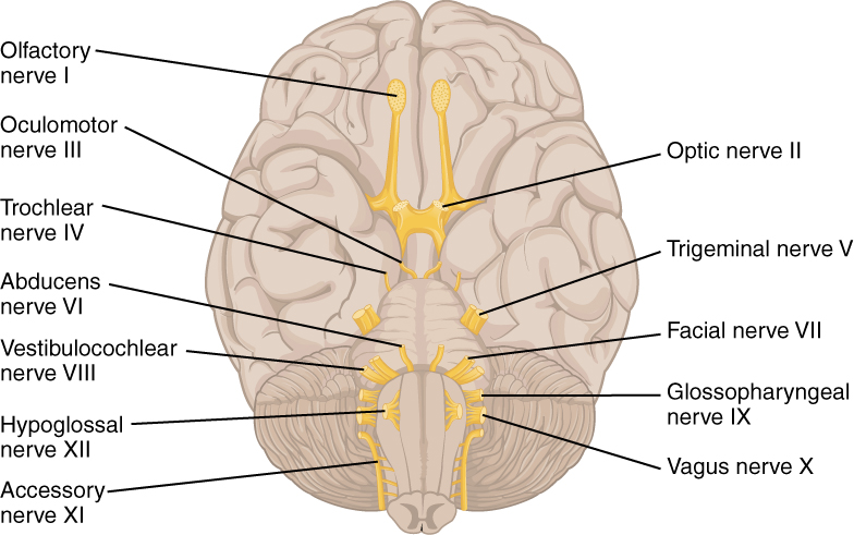
Focused cranial nerve assessment can include visual fields (II), eye movements (III, IV and VI), facial sensation and mastication (V), facial movement (VII), hearing and vestibular complaints (VIII), palate/voice/swallow-related observations (IX and X), shoulder shrug (XI) and tongue movement (XII). Smell (I) is not routinely needed in every visit.
New diplopia, dysphagia, dysarthria, asymmetric facial findings or acute cranial-nerve changes may indicate serious pathology and should be escalated in context. Use appropriate specialist referral for detailed swallowing and visual assessments.
Apply it to physical therapy
Start with the task complaint, observe movement, then identify the most informative impairment tests. Explain whether a finding is a symptom, observation or quantified measure. Review combination and timeline of deficits before drawing conclusions.
Practice the decision
A patient has poor target accuracy on finger-to-nose testing but normal manual strength and intact light touch. Is ‘weakness’ a sufficient explanation?
Reasoning: No. Examine timing, coordination, vision, proprioception and functional reaching. A coordination deficit can occur without prominent loss of strength.
Check your understanding
Which additional examinations help distinguish reduced proprioception from cerebellar incoordination?
Original lecture sources: 2 Exam & Eval.pdf; 11 & 12 Cranial Nerves.pdf; 8 PNS EMG NCV.pdf; 9 Spinal Cord.pdf.. Study note: One older cranial-nerve source has a sensory-nerve numbering typo. Use CN VIII for the vestibulocochlear nerve, and check your course material against current anatomy references. Independently reorganized for educational review; official test protocols and figures should be accessed from their original sources. Not an individual evaluation, diagnosis or treatment plan.


