
← Diagnostic Imaging class notes
FROM MY PT SCHOOL NOTES · DIAGNOSTIC IMAGING · UNIT 4 · CHAPTER 13
Chest and cardiopulmonary images
Review the relevant anatomy and imaging question before deciding what the finding may mean for rehabilitation.
Key imaging concepts
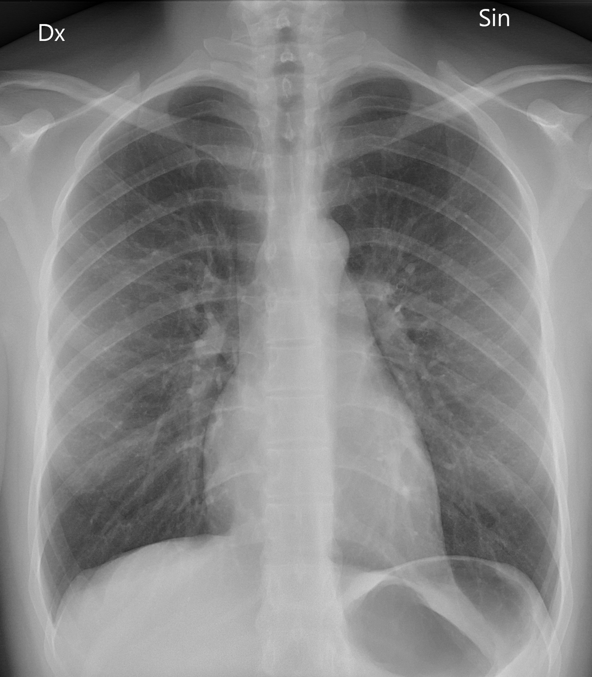
- Chest radiographs are read with a deliberate pattern: technical quality, airways, cardiac/mediastinal contour, diaphragm/pleura, lungs and bones.
- Check the study type (PA vs AP), rotation and inspiration; portable AP images can enlarge the apparent cardiac silhouette.
- An X-ray may aid a medical workup, but acute chest symptoms should be managed by presentation and urgency, not delayed awaiting imaging.
PT clinical connection
Chest pressure, severe dyspnea or acute hypoxia need immediate medical response.
High-yield distinction
Normal film is not a clearance test: Chest radiographic abnormalities may be subtle or time dependent; severe cardiopulmonary symptoms warrant emergency evaluation on clinical grounds. VERIFIED REAL-IMAGE REFERENCES (SOURCE LINKS ONLY) • Normal chest PA · CC0 1.0 · normal anatomy; not a pneumothorax • Pneumothorax · CC BY 4.0 · real radiographic case, multiple time points
Course source: 2 Cardiopulmonary.pdf; 1 Thoracic Spine.pdf. These are educational notes, not image reading certification or a substitute for an official report. Imaging orders and interpretation rights vary with professional scope and state law; image examples must be clinically verified and properly licensed.
← Unit 4 · ← Previous · Next →


