
FROM MY PT SCHOOL NOTES · NEUROLOGY · UNIT 1 · CHAPTER 02
Why do lesions produce different motor and sensory findings?
The site of a lesion changes what the therapist observes. Rather than memorizing a diagnosis from one sign, learn to distinguish central from peripheral patterns and connect a pathway to a functional problem.
Understand the essentials
UMN and LMN are different parts of the pathway
Upper motor neurons (UMNs) include descending motor systems that influence brainstem and spinal motor circuits. A lesion may lead to weakness, reduced selective control, and—over time—hyperreflexia, clonus or velocity-dependent resistance to stretch. Signs can evolve, especially after acute central injury.
Lower motor neurons (LMNs) directly innervate skeletal muscle from the anterior horn or brainstem motor nuclei. Damage commonly produces focal weakness, reduced tone and reflexes, atrophy and sometimes fasciculations. These findings are patterns, not stand-alone diagnoses: examination context and time course matter.
Sensory tracts carry different information
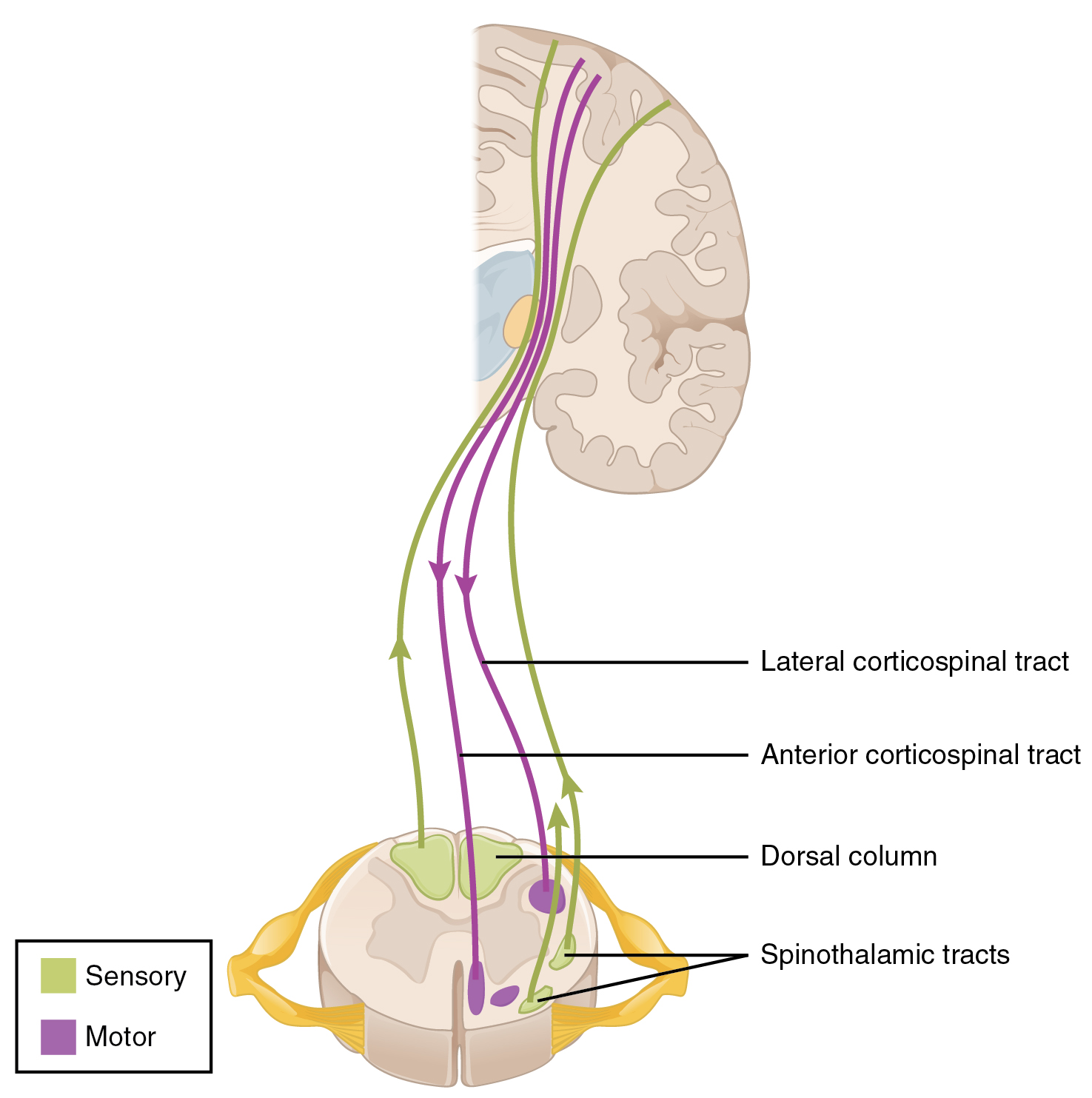
The dorsal column–medial lemniscus pathway contributes vibration, discriminative touch and conscious proprioception. The anterolateral system carries pain and temperature. Many dorsal-column signals ascend ipsilaterally within the cord before crossing in the medulla; many pain/temperature fibers cross in the cord over a short segmental distance.
The major lateral corticospinal system crosses predominantly at the caudal medulla, so a lesion above that crossing often affects opposite-side limb control, while an injury below it can affect same-side control. This simplification is only a starting point; multiple pathways and lesion extents can alter the pattern.
Interpret patterns, not isolated tests
Compare distribution, sidedness, reflexes, sensory modalities and function. For example, weakness with brisk reflexes and impaired fine motor control suggests a different process than weakness with diminished reflexes and nerve-distribution sensory loss.
A single intact modality or normal reflex cannot rule out central pathology. Sudden new unilateral weakness, speech changes or facial droop should prompt immediate emergency escalation rather than a lengthy outpatient localization exercise.
What changes for a physical therapist?
Use findings to test hypotheses: Is weakness isolated to one myotome, a peripheral nerve distribution, multiple limbs, or one body side? Is sensory loss modality-specific? What happens to gait, dexterity and postural control? Your goal is appropriate evaluation and referral, not claiming to localize a lesion from one sign.
Try the reasoning
A patient presents with gradually increasing hand clumsiness, gait difficulty and exaggerated leg reflexes. Should the therapist begin isolated hand-strengthening because the complaint is ‘hand weakness’?
Answer: No. The pattern raises concern for a central neurological process such as myelopathy and warrants timely medical assessment. Escalation urgency depends on progression and additional findings.
Check your understanding
Why might a unilateral spinal cord lesion affect same-side position sense but opposite-side pain and temperature below the lesion?
Original lecture sources: 16 Motor Pathways I.pdf; 16 Motor Pathways II.pdf; 16 Motor Pathways III.pdf; 9 Spinal Cord.pdf; 8 Sensory Receptor and the PNS.pdf.. Adapted into independent educational wording; diagrams and paid-companion cross-references are in preparation. Not a patient-specific diagnosis or care plan.


